What is Fluoroscopy?
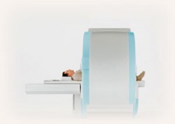
Fluoroscopy is the study of moving body structures. A continuous x-ray beam is passed through the body part being examined, it is then transmitted to a TV-like monitor so that the body part, and its motion, can be seen in detail. Fluoroscopy enables radiologists to look at and evaluate body systems including the digestive, reproductive, respiratory, skeletal, and urinary systems. Often times fluoroscopy is performed to evaluate specific regions of the body.
When is Fluoroscopy Used?
Fluoroscopy is used in many kinds of examinations such as arthrography, barium studies, and hysterosalpinography. Below is a list and brief description of the fluoroscopic exams offered at Advanced Medical Imaging.
Types of Fluoroscopic Exams and Preparation
Arthogram
An arthrogram is an exam in which contrast material is injected into the affected joint. Joint injections can be for an x-ray arthrogram, or simply used as a guide to insert contrast into the joint for an MRI Arthrogram. It is used to view the soft tissue structures of your joint (tendons, ligaments, muscles) that are not seen on a plain x-ray, and helps the radiologist evaluate the need for joint replacement procedures.
An arthrogram is performed to evaluate persistent, unexplained joint pain, swelling, or abnormal movement in the ankle, wrist, shoulder, hip, and knee.
How to Prepare for Your Arthrogram
- Please plan on arriving 15 minutes prior to your appointment for patient registration.
- Before arriving for your exam let the Scheduling Department at AMI know whether there is any chance you are pregnant or if you are allergic to iodine.
- Please do not eat or drink four (4) hours prior to your exam.
- Please make sure to bring a driver with you to take you home.
- Do not consume any aspirin products five (5) days prior to your exam.
Barium Enema
A barium enema is an examination of the large intestine. Barium enemas are used to help diagnose diseases and other problems that affect the large intestine such as colon cancer or colon polyps, unexplained weight loss, narrowing of the walls of the intestine. Barium enema’s can also be used to monitor the progression of diseases affecting the intestine. There are two types of procedures that can be performed: a single-contrast study, or an air-contrast study.
How to Prepare for Your Barium Enema Exam
- Please plan on arriving 15 minutes prior to your appointment for patient registration.
- Before arriving for your exam let the Scheduling Department at AMI know:
- Whether there is any chance you are pregnant.
- Are allergic to barium or latex.
- If you have had an upper digestive barium test (Upper GI or barium swallow) recently.
- 1-3 days before your exam please abide by a clear liquid diet.
- Day before test:
- Drink large amounts of non-carbonated clear liquids.
- Drink the prep material provided by AMI.
- Day of the test:
- Bowel preparation requires liquid passing through the bowel to be free of any stool.
- You may be required to repeat the enema until the liquid that passes is free of any stool.
Esophogram, Upper GI, and Small Bowel
These procedures examine the upper and middle portions of the gastrointestinal tract using contrast material, fluoroscopy, and x-ray. Just prior to the test you will swallow liquid barium and water. The radiologist then tracks the progress of the barium through your esophagus, stomach, and the first part of the small intestine.
These procedures are done for a variety of reasons, including:
- Determine whether chest pains are being caused by GERD.
- Determine whether the esophagus is functioning properly.
- Evaluate abdominal pain, difficulty swallowing, or vomiting.
- Evaluate weight loss and chronic diarrhea.
- Detect inflamed areas of the intestine.
- Detect ulcers, tumors, or polyps in the intestine.
- Detect blood in the stool.
- A history of Crohn's Disease.
How to Prepare for Your Esophogram, Upper GI and/or Small Bowel Exam
- Please plan on arriving 15 minutes prior to your appointment for patient registration.
- Please do not eat or drink anything after midnight prior to the day of your exam.
- Exam time varies from patient to patient based on the specific procedure being provided. Please inquire with the Scheduling Department.
Hysterosalpinogram
A hysterosalpinogram is a test that examines the inside of the uterus and the fallopian tubes and surrounding regions. It is often performed when a woman is unable to become pregnant or following a sterilization procedure. The pictures provided by the contrast material help reveal problems such as an injury or abnormal structure of the uterus or fallopian tubes, or blockage that would prevent an egg from passing through a fallopian tube.
How to Prepare for Your Hysterosalpinogram Exam
- Please plan on arriving 15 minutes prior to your appointment for patient registration.
- The exam will be scheduled 7-10 days after the first day of your menstrual period.
- Please abstain from sexual intercourse following the first day of your period until after the procedure.
- Please take a home pregnancy test the morning of your exam to confirm your pregnancy status.
Myelogram
Myelography is an examination of the spinal cord and the space around it. Myelograms render images that show distortions of the spinal cord, the spinal canal, and the spinal nerve roots. Myelograms are performed for the following reasons:
- To detect spinal lesions or other abnormalities.
- Ruptured or herniated disks.
- Bone spurs causing back pain due to pressure on nerves.
- Tumors in the spine or around the tissue.
- Infection, inflammation, abnormalities of blood vessels that supply the spinal cord, and other traumatic injuries.
How to Prepare for Your Myelogram Exam
- You will receive a letter in the mail prior to your procedure with more specific instructions regarding medications.
- Do not consume any NSAIDS and blood thinners five (5) days prior to your exam.
- Lab work is required prior to the exam. Office staff will contact you to speak further about this.
- Prior to your appointment you should let Advanced Medical Imaging scheduling department know if there is any chance you are pregnant or have an allergy to iodine.
- Please plan on arriving 15 minutes prior to your appointment for patient registration.
- Please do not eat or drink anything four (4) hours prior to your scheduled exam time.
- Please make sure to bring a driver with you to take you home.
- Please have a list of any medications you are currently taking ready for the technologist.
- Please remain on bed-rest 24 hours after your exam.
- Do not work for two (2) days following your exam.


